

A study led by researchers at Cedars-Sinai and NeuroVision Imaging LLC provides the scientific basis for using non-invasive eye imaging to detect the pathological hallmarks of Alzheimer’s.
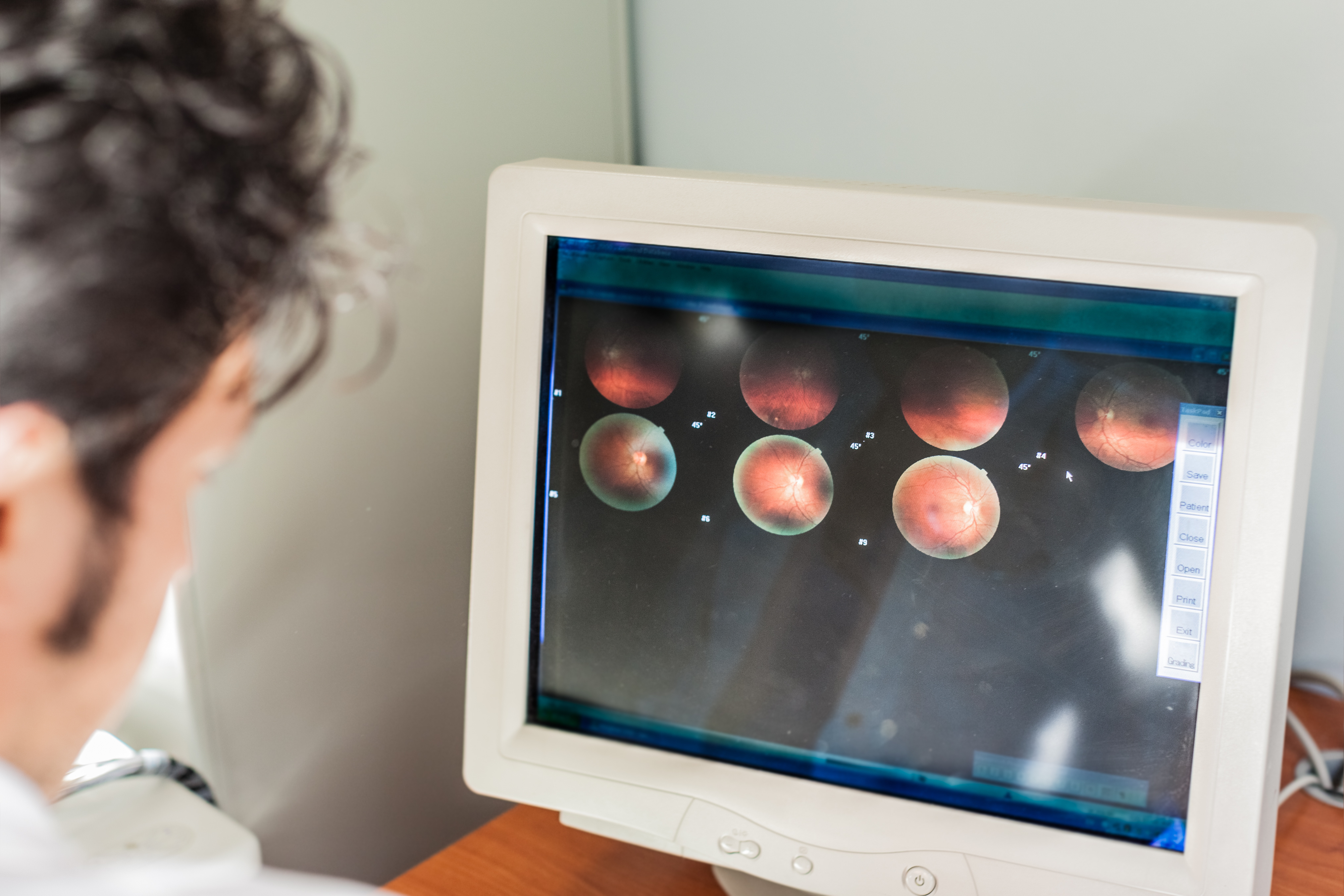
The experimental technology, developed by Cedars-Sinai and NeuroVision, scans the retina using techniques that can identify beta-amyloid protein deposits that mirror those in the brain.
Accumulations of neurotoxic beta-amyloid protein can be detected with positron emission tomography, or PET scans, and analysis of cerebrospinal fluid, but these are invasive, inconvenient, and costly, making them impractical for routine screening and follow-up evaluation.
‘This is the first study demonstrating the potential to image and quantify retinal findings related to beta-amyloid plaques non-invasively in living patients using a retinal scan with high resolution. This clinical trial is reinforced by an in-depth exploration of the accumulation of beta-amyloid in the retina of Alzheimer’s patients versus matched controls, and a comparison analysis between retina and brain pathologies.
‘Findings from this study strongly suggest that retinal imaging can serve as a surrogate biomarker to investigate and monitor Alzheimer’s disease,’ explained Maya Koronyo-Hamaoui, PhD, an Associate Professor of Neurosurgery and Biomedical Sciences and a research scientist at the Maxine Dunitz Neurosurgical Institute at Cedars-Sinai, and a co-founder, inventor and scientist at NeuroVision. She’s the senior leading author of an article in JCI Insight published online.
‘As a developmental outgrowth of the central nervous system that shares many of the brain’s characteristics, the retina may offer a unique opportunity for us to easily and conveniently detect and monitor Alzheimer’s disease,’ said Keith L Black, MD, Chairman of NeuroVision, Chair of the Department of Neurosurgery and Director of the Maxine Dunitz Neurosurgical Institute at Cedars-Sinai.
We know that Alzheimer’s begins as many as 10 or 20 years before cognitive decline becomes evident, and we believe that potential treatments may be more effective if they can be started early in the process. Therefore, screening and early detection may be crucial to our efforts to turn the tide against the growing threat of this devastating disease.’
Steven Verdooner, NeuroVision CEO, said that the imaging system leverages the company’s expertise in autofluorescence imaging of the retina using a specialised ophthalmic camera and sophisticated image processing software.
‘It’s exciting to see these studies demonstrating the power of the technology applied to the Alzheimer’s field. Our goal is to develop a product that is easy to use, affordable, and widely-accessible. We look forward to the potential of retinal imaging playing a vital role in solving the problem of Alzheimer’s, both in identifying and monitoring those who may be affected by the disease. Our next step is to continue with clinical trials, building upon the existing pharmaceutical company collaborations, to ensure our technology is ready for the medical community to help manage this disease.’


